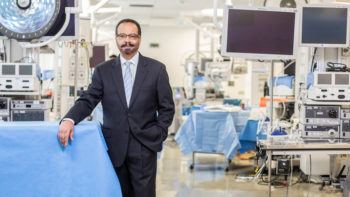
Groundbreaking New MRI Scanner Is The ‘Brainchild’ Of Imaging Pioneer, EnMed Dean Pettigrew
Researchers at Texas A&M are anxiously awaiting the spring delivery of an advanced generation MRI scanner that was inspired by the School of Engineering Medicine’s inaugural Dean Roderic Pettigrew.
Though health care providers use MRI scanners to diagnose, stage, monitor and research disease, the new technology is being developed to detect threats early and help prevent severe consequences, such as seeing coronary or vascular diseases before a heart attack or stroke, or identifying prostate cancer in its earliest stage before a biopsy.
Pettigrew — a former and founding director of NIH’s National Institute for Biomedical Imaging and Bioengineering — made the proposal in 2018 to “healthineers” at tech giant Siemens, challenging them to “push the limits of MRI performance physics to the furthest point that could be safely executed in the whole body.”
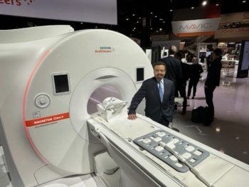
After four years in the development stage, Siemens unveiled the 3T Magnetom Cima.X2 in December at the annual meeting of the Radiological Society of North America in Chicago. The scanner offers unprecedented technical performance features when compared to conventional whole-body MRI systems in place now.
Pettigrew is working with Siemens on early scanning efforts to explore how best to utilize the unique technical features of the Cima, which is Spanish for the peak of a mountain. As a developmental scanner, the Cima is not yet commercially available. Pettigrew expects to receive the Cima scanner at Texas A&M’s EnMed Tower in Houston during Spring 2023.
“I realized that even modern MRI scanners had not yet been designed to reach the theoretical ‘summit’ of their performance capabilities,” said Pettigrew. “And that if we reach this summit, it could really improve our ability to identify early disease and prevent deleterious outcomes. Imagine that we could identify vulnerable plaque in arteries before it causes a heart attack or stroke, identify breast cancer even earlier than we can today and minimize the therapeutic intervention, or detect prostate cancer before biopsy to enable earlier treatment with noninvasive techniques.”
The science explained: Using a powerful magnet with various electromagnetic coils, an MRI works by temporarily magnetizing the body, sending a radiofrequency pulse into the area being imaged to stimulate the magnetized hydrogen in tissue molecules, then detecting the radiofrequency signal that the excited hydrogen nucleus returns. The signal is like one received by an FM radio. A computer processes the signal to make images for clinicians to use throughout biomedicine including neuroscience, musculoskeletal, oncology and cardiovascular applications. The ability to make an image and see fine detail depends on precisely controlled changes in the strength of the magnetic field in various directions. This is called a magnetic field gradient; the speed at which the change is made is called the slew rate.
What’s new: Siemens Cima offers a magnetic field gradient strength of 200 mT/m and a slew rate of 200 mT/m/s. Most conventional systems have a gradient strength for the whole-body only about 20% to 40% as strong as that of the Cima. In an MRI scanner, the amplitude of the magnetic field gradient is a major determinate of the smallest size structure that can be seen, and its sensitivity to motion and water diffusion, while the slew rate, or speed at which the gradient coils pulse, determines the speed of signal formation. As explained by Pettigrew, this is important for detecting things that happen quickly, like coronary motion.
Pettigrew said the strong gradients can also make images of microstructures like cardiac fibers clearer. As a result, the Cima could be a powerful tool to help clinicians and scientists better understand and treat highly prevalent problems such as prostate cancer and early-stage cardiovascular disease.
“This is an exciting development in the field of whole-body MRI, and its potential to improve clinical care and research is just beginning to be explored,” Pettigrew said. “Achieving such an advanced MRI system required a fresh design of the whole-body scanner and even new tools to build it. This scanner is unique in the world. We hope it will bring new understanding, greater prevention, and improved treatment of human disease.”
Pettigrew’s work on the Cima was sponsored by a Governor’s University Research Initiative (GURI) award. Established in 2015, GURI aims to attract to Texas universities transformative researchers who will catalyze the Texas economy into the future.