New Method Allows Scientists To Quickly Identify Individual Virus Particles
Influenza, SARS-CoV-2 and other viruses come in a wide variety of shapes and sizes, and by studying these shapes, scientists can learn how they function and how viral illnesses might be conquered.
Now, a team of Texas A&M AgriLife researchers has demonstrated a noninvasive way to study viruses that is quicker and more detailed than the current “gold standard” method. The study appeared recently in Analytical Chemistry.
“The need for very fast and accurate virus identification has always been important in the past, and this year it’s even more important because we know that viruses change; they mutate,” said Dmitry Kurouski, assistant professor in the Texas A&M University College of Agriculture and Life Sciences Department of Biochemistry and Biophysics, who led the study. “If someone has flu-like symptoms, how can we quickly distinguish the flu from COVID-19?”
Most viruses are too small to be seen under a typical microscope, so scientists often study flash-frozen virus samples with electron microscopes. These tools use beams of electrons to probe the virions’ intricate molecular structures. However, preparing samples for electron microscopy is time- and labor-consuming.
Kurouski’s team used a combination of two sophisticated techniques that, in theory, should require almost no sample preparation. Kurouski’s lab is unique in its ability to use both methods — tip-enhanced Raman spectroscopy and atomic-force microscopy–infrared spectroscopy. Tianyi Dou, a graduate student in Kurouski’s lab, performed the experiments.
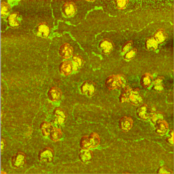
In both methods, the samples are approached with an extremely sharp metal needle covered with gold. Tip-enhanced Raman spectroscopy detects how a sample scatters laser light. This technique enabled the team to determine the viruses’ overall build and surface qualities and composition.
Using atomic-force microscopy–infrared microscopy, the team was able to see how the samples absorb infrared light. This allowed the researchers to gain information about the virions’ internal structure.
By combining the two methods, the team compared the surface and overall structure of the virus that causes cold sores, herpes simplex virus Type 1, to that of a virus that infects bacteria, bacteriophage MS2. The researchers were able to distinguish the herpes and bacteriophage virions with 100% accuracy.
Furthermore, the results were consistent with those obtained by the gold standard method, cryo-electron microscopy. Junjie Zhang, assistant professor in the Department of Biochemistry and Biophysics, led the electron microscopy experiments.
Besides the need to prepare and freeze samples, the gold-standard method has another drawback, Kurouski said. Cryo-electron microscopy involves averaging shapes to build a three-dimensional model of a representative virus. However, this averaging can conceal the true range of shapes a virus can take.
“Viruses can be described in terms of a Lego puzzle. Different structures can be built from the same building blocks,” Kurouski said.
And, a virus’s shape likely plays a big part in infection, he said, because the first step toward infection occurs at the surfaces of the virus and the host cell.
The new combination of methods does not average shapes but instead examines individual virus particles.
“Before, we had no tool to study this heterogeneity,” he said. “Now, we can look at what actually happens in nature.”
This article by Olga Kuchment originally appeared on AgriLife Today.