Werner The Wonder Dog
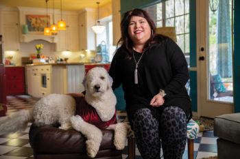
When Andrea Streicher saw the photos of the curly-haired, golden puppies a friend had sent her, it was love at first sight. Her day had started off with a series of unfortunate events — including a flat tire — but there in front of her was the antidote to all her doldrums.
Werner, a “double doodle” with Goldendoodle and Labradoodle parents, immediately joined the Streicher household and proved himself to be a happy, social and active dog.
He could often be found running around a dog park in their hometown of Austin, donning silly costumes or funky bandannas that matched his personality. Because of Werner’s excellent demeanor with children, Streicher aspired to train him as a therapy dog.
“We dyed his ears blue for his first birthday, and I noticed when his ears were blue that little kids were so drawn to him,” Streicher said. “We now keep his ears blue, so that when he goes to schools to help with readings and stuff, the kids will still be drawn to him.”
A DISHEARTENING DIAGNOSIS
At just around 2 years of age, Werner started becoming lethargic and depressed and began having seizures.
Concerned, Streicher took him in to her local veterinarian, where they found that Werner had a huge, rapidly growing skull tumor.
“When we first started having problems, the tumor was probably the size of my thumbnail. It was like a grape,” Streicher recalled. “Three weeks later, it was almost the size of an egg.”
Streicher met with several veterinarians who told her they had never seen such an aggressive tumor in such a young dog, and they feared his tumor would be inoperable.
The diagnosis came as a shock for Streicher and her family.
“We waited 24 hours until I could talk with the neurologist, and she basically said that it was huge,” she said. “It was super invasive. She said our main objective should be to keep Werner comfortable.”
Despite the grim prognosis, Streicher wasn’t about to give up on Werner, so she decided to take him to Texas A&M’s Veterinary Medical Teaching Hospital (VMTH).
“I thought he wasn’t going to survive, and if I lost him, I wanted his body to go to the veterinary students at Texas A&M so that they could study him and learn from his disease,” Streicher said.
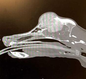
There, the VMTH team swiftly admitted Werner for a CT scan and confirmed the tumor took up a significant portion of his skull and was compressing his brain.
After the veterinary team spent some time analyzing Werner’s case, they gave Streicher some news she didn’t anticipate: They would be able to operate and attempt to remove the tumor. Werner would likely lose an eye and there was no guarantee of a successful surgery, but the doctors were confident they could give Werner’s case a shot.
The next step before surgery was to perform a bone biopsy to confirm the specific type of cancer Werner was suspected to have. After about two weeks of waiting, the biopsy results were returned and confirmed that Werner’s tumor was a multilobular tumor of bone.
Multilobular tumors are rare, and there are no known predispositions to this particular type of cancer. The tumors typically occur on flat bones, and in canines, the skull is the most common area they arise. Surgical removal of these tumors is tricky, and it is often impossible to remove the tumors from the skull with clean margins.
COLLABORATING TO CURE
On Oct. 10, 2018, when Werner checked in for his surgery, the VMTH neurology and oncology teams had come up with an innovative solution.
Because of the amount of skull Werner would lose in the operation, his clinicians would need to find a way to rebuild his skull from scratch.
Dr. Michael Deveau, a clinical assistant professor in the Texas A&M College of Veterinary Medicine & Biomedical Sciences (CVM) and holder of the Katherine and Rebecca Rochelle Chair in Oncology, reviewed CT scans of healthy dogs to generate a new skull piece that would replace the excised portion of bone. He collaborated with Dr. Elizabeth Scallan, in the Clinical Skills Laboratory, to make a 3D print of the skull replacement pieces and to engineer a mold that could be used in the operating room.
During surgery, Dr. Joseph Mankin, a clinical associate professor in the neurology service, and Dr. Maya Krasnow, a second-year resident in neurology, filled the mold with a material called poly methyl methacrylate (PMMA), a type of shatter-resistant plastic.
The PMMA hardened in minutes, and readily created Werner’s new skull. Beside them, Dr. Brandon Wustefeld-Janssens, one of the VMTH’s veterinary surgical oncologists, removed Werner’s right eye.
After his eight-hour surgery, Werner faced a long recovery.
“The first night was rough,” Streicher remembered. “I’m not going to lie — I wondered if I did the right thing, if I kept him here for me or if I kept him here because it was the right thing to do.”
But after several nights of sleeping by Werner’s side at home, things started to look up. Werner’s original, playful attitude started to shine through again, so much so that he had to return to the VMTH mid-recovery to have more stitches placed around his incision areas for security.
“I literally had to hide all the toys, because we wouldn’t throw them to him, so he would throw the toys himself,” Streicher said with a laugh.
Werner’s team is equally impressed with his recovery and credit collaboration for the success of his operation.
“When a client comes to an academic institution like the Texas A&M Veterinary Medical Teaching Hospital, they’re typically coming because we offer strengths that other practices may not be able to provide,” Deveau said. “We should be able to do things that nobody else can do, and if there isn’t an easily apparent way to do something, then we can pool our resources, put our heads together, and have an outcome that may benefit patients like Werner.”
Streicher hopes to have Werner certified as a therapy dog to help comfort children undergoing cancer treatment.
“He is just amazing,” Streicher said. “The fact that he’s still here is just a miracle. He is Werner the Wonder Dog.”
The Streicher family is grateful for the efforts made by the VMTH team to save Werner’s life. Specifically, Streicher said she credits Texas A&M with “taking the chance.” For that, she says she will always recommend the VMTH for quality veterinary care.
“If you need any care that goes above and beyond, just go to Texas A&M. Just do it. You won’t be sorry,” she said.
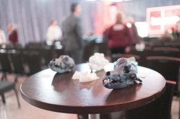
At the end of February, Streicher brought Werner back to College Station, but as a guest of honor instead of a patient.
Werner’s story was showcased as one of four exhibits at the Texas A&M Foundation’s Exploration Day, which provides immersive experiences from disciplines across campus for top university donors.
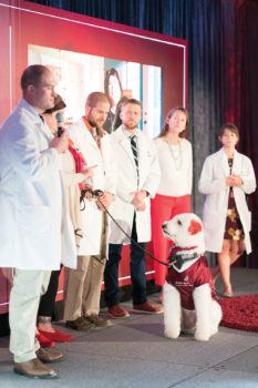
Werner appeared on stage as the grand finale of the college’s exhibit and received a standing ovation.
As a tribute to the team that saved his life, Werner wore a 12th Man jersey, while his famous blue ears were dyed maroon to honor the Aggies who never gave up on saving his life.
This article by Corley-Ann Parker originally appeared in CVM Today.