Think of them as nature’s molecular Swiss army knife.
Iron-sulfur clusters are tiny metallic ions found in all living organisms that perform essential biological tasks, from respiration in humans to photosynthesis in plants. Now, a Texas A&M University-led study is providing new insight into how they function.
For the first time ever, chemists at Texas A&M, the Massachusetts Institute of Technology and the University of Utah have mapped the structure of the cysteine desulfurase complex in eukaryotic systems, an essential three-protein framework largely responsible for the biosynthesis of iron-sulfur clusters. Its composition reveals the ways the proteins interact, an important clue in understanding the process of how they come together to generate an iron-sulfur cluster and deliver it to its respective target in the cell.
Their findings are published today (Jun. 20) in the current issue of the Proceedings of the National Academy of Sciences (PNAS) Online Early Edition. The work was funded in part by the National Institutes of Health and the National Science Foundation.
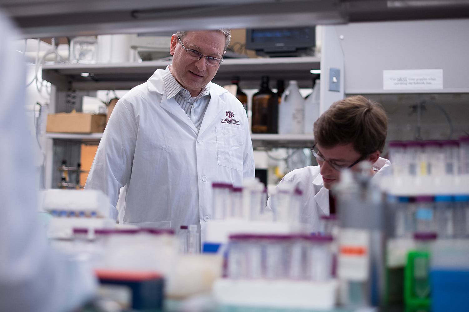
“Iron-sulfur clusters are ancient protein cofactors that are required by many enzymes in essential cellular processes,” said Texas A&M chemist and principal investigator David Barondeau. “There are literally hundreds of different human proteins that require iron-sulfur clusters, and yet, we don’t really understand fundamental details for how the biosynthetic machinery makes these cofactors.”
Iron-sulfur cluster biosynthesis is an evolutionarily conserved pathway, meaning that it occurs in both eukaryotes (animals and plants) and prokaryotes (bacteria and archaea). However, previous studies have demonstrated a stark difference in how it occurs within the two systems. While the cysteine desulfurase complex forms the core of the iron-sulfur assembly complex in eukaryotic systems, two of its proteins — ISD11 and ACP — are not found in the prokaryotic process.
Further perplexing scientists is the way a protein called frataxin (FXN) in eukaryotes binds to the cysteine desulfurase complex and accelerates the rate of iron-sulfur cluster biosynthesis, whereas in prokaryotes, its structural counterpart, CyaY, actually inhibits iron-sulfur cluster assembly. The Barondeau Laboratory wanted to find out why.
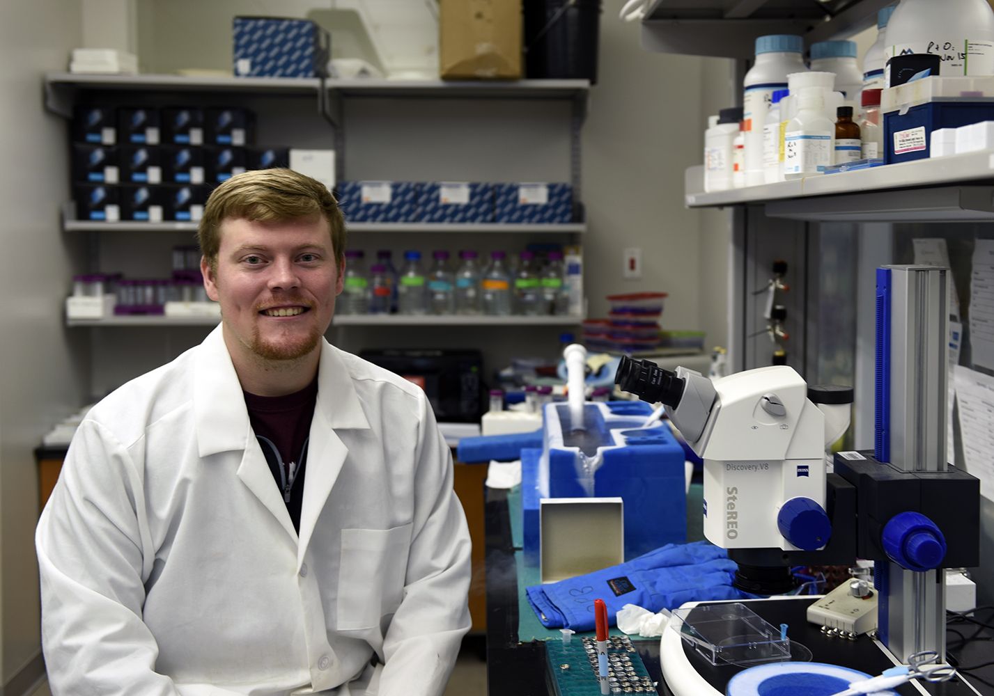
Chemistry graduate student Seth Cory ’18, primary scientist for the study and first author on the resulting paper, says that understanding the architecture of the cysteine desulfurase complex will lay the groundwork to help answer these and other questions about how iron-sulfur clusters are built.
“We took all of our tools and threw it at this system to figure out how the proteins interact with one another,” said Cory, a member of the Barondeau Lab since June 2014. “It was nice to see all these things fit together. It was a nice biochemical and biophysical approach.”
To determine the structure of the cysteine desulfurase complex, the researchers used X-ray crystallography. After crystallizing the protein complex, the crystals were frozen in liquid nitrogen and sent to the Stanford Synchrotron Radiation Lightsource (SSRL) facility. There, they were exposed to high-intensity X-ray beams to obtain diffraction data that describes the structure of the protein complex in the crystal. Interactions observed in the crystal structure then were perturbed by making mutations in the analogous yeast proteins. The effect of these mutations in yeast allowed for the evaluation of protein-protein interfaces observed in the crystal structure to be investigated in an in vivo setting. Finally, an electron-density map of the complex was generated using negative stain electron microscopy, which also corresponded with the crystal structure data.
The team’s studies support a novel cysteine desulfurase architecture that Barondeau says provides clues to how eukaryotic mitochondria control the activity of this important biosynthetic pathway.
Beyond satisfying basic curiosity and pushing the boundaries of biochemistry, Cory says the results also have potential medical applications. Scientists in recent years have become increasingly interested in the frataxin protein and its ties to a rare neurological condition called Friedreich’s ataxia (FRDA) that affects one in 50,000 people.
FRDA patients possess a genetic mutation that prevents adequate production of the frataxin protein. Currently, there is no cure, but Cory says understanding the mechanism of iron-sulfur cluster biosynthesis may one day lead to viable treatment options.
“Friedreich’s ataxia leads to symptoms such as loss of coordination, cardiomyopathy and aggressive scoliosis — all which diminish patients’ quality of life,” Cory said. “I think the biggest thing that will come out of our current results is that we now have a platform for future studies. We have a platform to design other experiments to test the function of frataxin in the context of the three-protein complex. That’s really the biggest and most controversial question in the field: How does frataxin function?”
Although their results reveal vital information about an important protein complex, Cory admits they simultaneously present many new questions. For him, one of the most important takeaways is recognition of the critical role multidisciplinary research plays in the advancement of science.
“Not only are protein structures cool, but we can learn a lot from them,” Cory said. “Collaborative research is really important for advancing our science every day. If we can get a great group of people together, we can get it done. I think that’s what we’ve demonstrated here.”
The team’s PNAS paper, “Structure of human Fe-S assembly sub-complex reveals unexpected cysteine desulfurase architecture and acyl-ACP-ISD11 interactions,” can be viewed online along with related figures and captions.
###
This story by Chris Jarvis originally appeared on the College of Science website.